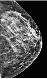
When a radiologist looks at your mammogram, she or he is looking for anything that looks out of the ordinary, such as asymmetry, irregular density, or thickening skin.[1] For this reason, it is very helpful to have past breast images available to your radiologist for historical comparison. Suspicious features often show up as shadows on a mammogram that need further investigation, often by ultrasound, to better understand their nature.[2] When radiologists examine a structure in the breast closely, they look for specific characteristics that can help determine the likelihood that the structure is malignant.
Is it solid or fluid-filled?
 Fluid-filled simple cysts are benign and very common in women, particularly between the ages of 30 and 50. An ultrasound exam can help to determine whether a round mass is solid or filled with fluid, a sign that it is a simple cyst. but it is not a perfect tool for this purpose. A mass can also appear solid when it consists mostly of fluid with small solid particles suspended in it. Even if the mass is only partially solid, however, further follow-up tests will likely be recommended. It’s important to keep in mind, however that solidity does not equal malignancy. Some conditions, such as benign fibroadenomas (the most common form of breast mass), appear as solid masses.[3]
What shape does it take?
A mass is more suspicious when it has ill-defined, spiked, or blurry borders, which can indicate infiltration of the surrounding tissue. Furthermore, a round or oval mass is more indicative of a benign condition than one with an irregular or lobule-like shape.[4]
How dense is it?
The density of a mass is inversely related to the amount of fatty tissue it contains. If one region of the breast contains a significantly lower proportion of fatty tissue than the surrounding area, it is considered high density and more likely to contain cancer cells. Bear in mind, however, that breast density varies greatly from one woman to the next, such that normal density for one woman may represent a red flag for another. What the radiologist is looking for is change and inconsistency. Even if your mammogram shows change over time or a variation in density, it is not immediate cause for alarm; a number of benign causes, including normal variations, trauma, and diabetic mastopathy, are possible.[5]
What form do any calcifications take?
Calcifications, small calcium deposits that develop in the breast, are very common findings on mammograms. They can take the form of either macrocalcifications, which appear as large white dots on the mammogram, or as microcalcifications, which appear as tiny white flecks. Macrocalcifications are almost always noncancerous and require no further testing. Microcalcifications are of concern when they appear as clusters, which can be a sign of ductal carcinoma in situ (DCIS), a non-invasive form of breast cancer.[6]
If your breast imaging shows abnormalities that are suspicious for breast cancer, then the radiologist will recommend a biopsy. It’s important to remember that breast biopsies come back negative around 80% of the time, so even if your doctor recommends this test, it does not necessarily mean that you are likely to have cancer.
To learn more about your mammogram and what to expect, download our free ebook.
 The information contained in the Iowa Radiology website is presented as public service information only. It is not intended to be nor is it a substitute for professional medical advice. You should always seek the advice of your physician or other qualified healthcare provider if you think you may have a medical problem before starting any new treatment, or if you have any questions regarding your medical condition.
Iowa Radiology occasionally supplies links to other web sites as a service to its readers and is not in any way responsible for information provided by other organizations.
Sources
[1] http://www.breastcancer.org/symptoms/testing/types/mammograms/interpret
[2] http://breast-cancer.ca/mass-characteristics/
[3] http://www.webmd.com/breast-cancer/benign-breast-lumps
[4] http://breast-cancer.ca/mass-chars/
[5] http://radiopaedia.org/articles/asymmetrical-density-in-mammography
[6] http://www.imaginis.com/mammography/diagnostic-mammography

