Ultrasounds are a safe, easy way to monitor fetal development, examine ovarian abnormalities, investigate potential causes of infertility, or identify cancerous and benign tumors. Depending on the purpose of your women's imaging procedure, your doctor might recommend that you have a transvaginal rather than a traditional transabdominal pelvic ultrasound. Transvaginal ultrasounds can allow your care providers to make a much more accurate diagnosis or care plan and can prevent serious complications during pregnancy, including dramatically lowering the risk of pre-term labor.
What happens during a traditional ultrasound?
An internal ultrasound is a way to peek beneath your skin, like an x-ray but without the dangers that ionizing radiation can pose. It can also show live, real-time pictures of what’s happening beneath the surface rather than just a static image.
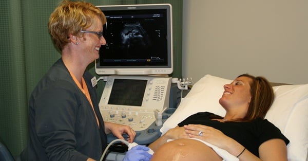
In an ultrasound[1], a technologist applies a lubricant gel to your skin to reduce friction and help the sensor move around easily. The sensor (called a “transducer”) emits high-frequency sound waves, which travel into your body and bounce off your internal organs and structures at different speeds. A computer reads the pattern of the sound waves as they echo back to the sensor and translates them into a real-time visual representation of the internal structures.
Other Types of Ultrasounds
Transabdominal Ultrasounds
In a transabdominal ultrasound, the technologist moves the transducer over the surface of your abdomen, which shows a general picture of your abdominal structures. In a transvaginal ultrasound[2], a wand-shaped transducer is inserted into the vagina. The sensor is covered with a probe cover and lubricant and should not cause much discomfort. By inserting the sensor into the vagina, the technologist can get closer and more detailed images of many of the organs and internal structures of the abdomen and reproductive system, allowing for a more accurate evaluation by the radiologist. It also enables a technologist to detect and hear a fetal heartbeat much earlier in a pregnancy than a transabdominal ultrasound and to look at a developing fetus in much greater detail.
Transvaginal Ultrasounds
Recent scientific studies have recommended transvaginal ultrasound as the routine mid-trimester obstetric ultrasound scan. Unlike transabdominal ultrasounds, transvaginal ultrasound can detect some cervical changes that could indicate complications, such as a likelihood to deliver pre-term, early enough to potentially prevent problems.[3] A transvaginal ultrasound can reveal whether a woman’s cervix is shortening an abnormal amount (1.5 cm or shorter in a low-risk patient, or 2.5 cm in a high-risk patient), which indicates a much greater risk of premature delivery. This shortening begins to occur weeks before other indicators of preterm labor and can result in a myriad of developmental problems and lowered chance of newborn survival. If the shortening is detected early enough, doctors can begin prescribing progesterone to help prevent the onset of preterm labor and its associated consequences.[4]
Since transvaginal ultrasounds are quite safe, the benefits of evaluating whether these abnormalities exist greatly outweigh any potential drawbacks. It has been recommended that pregnant women receive, as a matter of course, a transvaginal ultrasound to measure their cervical length sometime between 18 and 24 weeks of gestation.[5]
In addition to being a valuable screening tool during pregnancy, transvaginal ultrasounds allow a technologist to safely examine other reproductive system issues, such as cysts, fibroid tumors, or other growths; abnormal vaginal bleeding and menstrual problems; certain types of infertility; ectopic pregnancy; or other pelvic pain.[6]
To learn more about your ultrasound options and get information about other health care procedures, subscribe to our blog.
The information contained in the Iowa Radiology website is presented as public service information only. It is not intended to be nor is it a substitute for professional medical advice. You should always seek the advice of your physician or other qualified healthcare provider if you think you may have a medical problem before starting any new treatment, or if you have any questions regarding your medical condition.
Iowa Radiology occasionally supplies links to other web sites as a service to its readers and is not in any way responsible for information provided by other organizations.
[1] https://www.radiologyinfo.org/en/info.cfm?pg=genus#:~:text=In%20an%20ultrasound%20exam%2C%20a,sound%20waves%20into%20the%20body.
[2] https://www.healthline.com/health/transvaginal-ultrasound#procedure
[3] Campbell, Stuart. “Universal cervical-length screening and vaginal progesterone prevents early preterm births, reduces neonatal morbidity and is cost saving; doing nothing is no longer an option.” Ultrasound Obstet Gynecol 38:1-9. ISUOG, 2011.
[4] Ibid.
[5] Werner, E.F. et al. “Universal cervical-length screening to prevent preterm birth: a cost-effectiveness analysis.” Ultrasound Obstet Gynecol 38:32-37. ISUOG, 2011.
[6] https://www.hopkinsmedicine.org/health/treatment-tests-and-therapies/pelvic-ultrasound


